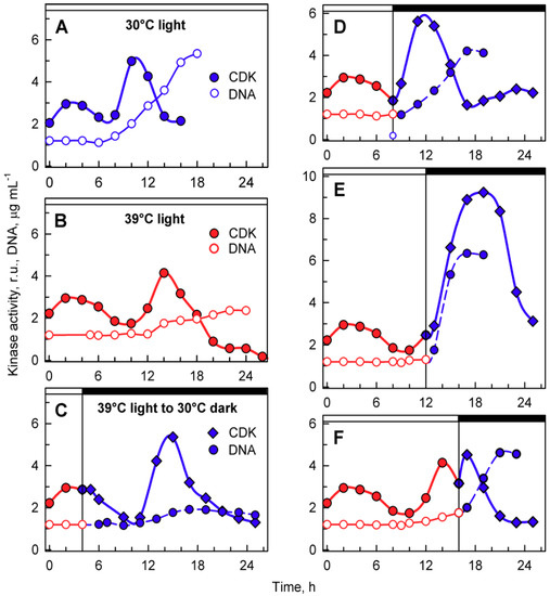
38 chlamydomonas diagram with labels
Structure of Chlamydomonas (With Diagram) | Genetics In this article we will discuss about the structure of chlamydomonas (explained with labelled diagram). The unicellular green alga Chlamydomonas is haploid with a single nucleus, a chloroplast and several mitochondria (Fig. 9.3). It can reproduce asexually as well as sexually by fusion of gametes of opposite mating types (mt + and mt - ). Structure and Diagram of Volvox and Their Functions - NotesHippo Volvox Structure: Diagram of Volvox with Label The cells of anterior end possess bigger eye spots than those of posterior end cells. The cells of posterior side become reproductive on maturity. Thus, spherical or round colony of Volvox shows clear polarity. Cell structure of volvox colony are Chlamydomonas type.
Diagram of Chlamydomonas angulosa... - Getty Images UNSPECIFIED - CIRCA 2003: Diagram of Chlamydomonas angulosa, Flagellated Protozoan. Drawing. (Photo by DeAgostini/Getty Images)

Chlamydomonas diagram with labels
Use this labeled diagram of a chlamydomonas cell to - Course Hero Use this labeled diagram of a Chlamydomonas cell to address the following two questions. 32. Which of the following statements correctly identifies aspects related to photosynthesis and/or respiration? 1. Acetyl CoA is most often found in G. 2. NADPH accumulates in C. 3. ATP is found in F. 4. Morphology of Chlamydomonas (With Diagram) | Algae - Biology Discussion In this article we will discuss about the external morphology of chlamydomonas. Also learn about its Neuromotor Apparatus, Electron Micrograph, Palmella-Stage with suitable diagram. 1. The organism is an unicellular alga (Fig. 11). 2. The thallus is spherical to oblong in shape but some species are pyriform or ovoid. ADVERTISEMENTS: 3. Labeled Diagram of Spirogyra - QS Study Labeled Diagram of Spirogyra. Plant kingdom. Spirogyra is a sophisticated, filamentous green alga, found in freshwater represented by about 300 species. It is also identified as pond silk, as its fiber burnishes like silk due to the occurrence of mucilage.
Chlamydomonas diagram with labels. Eye Diagram With Labels and detailed description - BYJUS A brief description of the eye along with a well-labelled diagram is given below for reference. Well-Labelled Diagram of Eye The anterior chamber of the eye is the space between the cornea and the iris and is filled with a lubricating fluid, aqueous humour. The vascular layer of the eye, known as the choroid contains the connective tissue. Chloroplast Structure and Function in detail with Labelled Diagram The chloroplasts are the cell organelles which consist of these pigments. The 3 types of pigments present in plants are chlorophyll, carotenoids, and anthocyanins. Chlorophyll imparts the green color to plants. Plastids are membrane-bound cytoplasmic organelles that can be found in the cells of plants and algae. Spirogyra Labelled Diagram Spirogyra (common names include water silk, mermaid's tresses, and blanket weed) is a genus of filamentous charophyte green algae of the order Zygnematales, named for the helical or spiral arrangement of the chloroplasts that is characteristic of the genus. Draw a labelled diagram of Spirogyra. 51 Differentiate between flying lizard and bird. Chlamydomonas as a Model Organism - Rice University Chlamydomonas as a Model Organism. Chlamydomonas, a genus of unicellular photosynthetic flagellates, is an important model for studies of such fundamental processes as photosynthesis, motility, responses to stimuli such as light, and cell-cell recognition.C. reinhardi, the most commonly studied species of Chlamydomonas, has a relatively simple genome, which has been sequenced.
Problem 24TY from Chapter 21 - Chegg LearnSmart Online for Biology | 10th Edition. ISBN-13: 9780077350611 ISBN: 0077350611 Authors: Sylvia S Mader Rent | Buy. This is an alternate ISBN. View the primary ISBN for: Biology 10th Edition Textbook Solutions. Microtubules filaments of the cytoskeleton: A) Chlamydomonas ... Download scientific diagram | Microtubules filaments of the cytoskeleton: A) Chlamydomonas reinhardtii fluorescently labeled with an antibody to tyrosinated tubulin (©2018 Courtesy of Karl ... Genetic map of the Chlamydomonas reinhardtii plastid genome.... Download scientific diagram | Genetic map of the Chlamydomonas reinhardtii plastid genome. Protein-coding regions are yellow and their exons are labeled with an "E" followed by a number denoting ... Life Cycle of Chlamydomonas (With Diagram) - Biology Discussion Each daughter cell develops cell wall, flagella and transforms into zoospore (Fig. 6). The zoospores are liberated from the parent cell or zoosporangium by gelatinization or rupture of the cell wall. The zoospores are identical to the parent cell in structure but smaller in size. The zoospores simply enlarge to become mature Chlamydomonas.
LABORATORY 9 - Susquehanna University Labeled diagram of Chlamydomonas. ... Chlamydomonas from culture. Cells have been stained with Lugol's Iodine, which complexes with true starch to turn black. 400X . You have slides of colonial volvocine green algae, which include Volvox, Gonium , Eudorina, ... Animal Cells: Labelled Diagram, Definitions, and Structure - Research Tweet Only present in lower plant forms (e.g. chlamydomonas) Present in all animal cells: Chloroplast: Plant cells have chloroplasts to synthesize their own food. Absent: Plasma Membrane: Cell wall and a cell membrane: Only cell membrane: Flagella: Present in some cells (e.g. sperm of bryophytes and pteridophytes, cycads and Ginkgo) Chlamydomonas: Position, Occurrence and Structure (With Diagrams) Chlamydomonas is unicellular, motile green algae. The thallus is represented by a single cell. It is about 20 p,-30|i in length and 20 µ in diameter. The shape of thallus can be oval, spherical, oblong, ellipsoidal or pyriform. The pyriform or pear shaped thalli are common, they have narrow anterior end and a broad posterior end (Fig. 1). Chlamydomonas - Wikipedia Chlamydomonas globosa, again with two flagella just visible at bottom left Chlamydomonas is a genus of green algae consisting of about 150 species [2] all unicellular flagellates, found in stagnant water and on damp soil, in freshwater, seawater, and even in snow as "snow algae". [3]
Indian Botanists: Algae may be heterotrophic!
Describe the structure of chlamydomonas with neat labelled diagram ... answeredOct 30, 2020by Naaji(56.8kpoints) selectedOct 30, 2020by Jaini Best answer 1. Chlamydomonas is a simple, unicellular, motile fresh water algae. They are oval, spherical or pyriform in shape. 2. The cell is surrounded by a thin and firm cell wall made of cellulose. 3. The cytoplasm is seen in between the cell membrane and the chloroplast. 4.

Flashcards Table on Biology 121 Lab Exam 1
Chlamydomonas reinhardtii - an overview | ScienceDirect Topics Chlamydomonas reinhardtii cells are oval shaped, c. 10 μm in length and 3 μm in width, with two flagellae at their anterior end ( Figure 1 ). The cells contain a single chloroplast occupying 40% of the cell volume and several mitochondria. These cells exist as mating-type (+) or mating-type (-).

Chlamydomonas
Structure of Chlamydomonas (With Diagram) | Chlorophyta In this article we will discuss about the structure of chlamydomonas with the help of suitable diagrams. Chlamydomonas is unicellular, motile green algae. The thallus is represented by a single cell. It is about 20 p,-30|i in length and 20 µ in diameter. The shape of thallus can be oval, spherical, oblong, ellipsoidal or pyriform.

ODA10 localizes to a unique proximal flagellar domain. (A) IF... | Download Scientific Diagram
Biological drawings. Structure of Chlamydomonas. Learning Resources for ... Structure of Chlamydomonas: Next Drawing > Chlamydomonas is the name given to a genus of microscopic, unicellular green plants (algae) which live in fresh water. Typically their single-cell body is approximately spherical, about 0.02 mm across, with a cell wall surrounding the cytoplasm and a central nucleus.
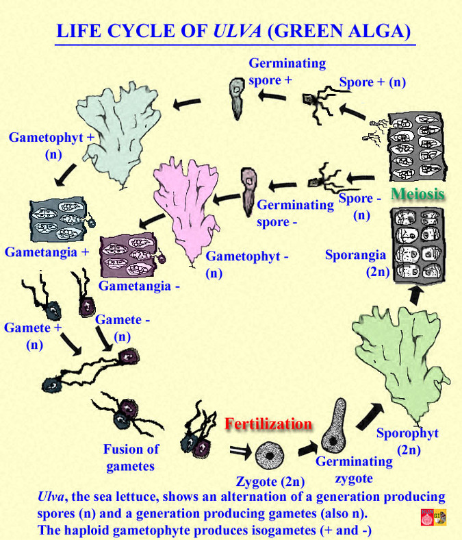
Sea Lettuce - Living Marine Resources BIO 340 - Marine Biology Columbia College SC Spring 2013
Draw a neat labelled diagram. Chlamydomonas - Biology Draw a neat labelled diagram. Chlamydomonas . Maharashtra State Board HSC Science (General) 11th. Textbook Solutions 8018. Important Solutions 19. Question Bank Solutions 5546. Concept Notes & Videos 439. Syllabus. Advertisement Remove all ads. Draw a neat labelled diagram. ...
37+ Ideas Draw A Labelled Diagram Of Amoeba Paramecium And Penicillium - Label Template Online
Amoeba Diagram Illustrations, Royalty-Free Vector Graphics ... - iStock Cross section of a Chlamydomonas. Structure of the algae cell. Vector diagram for educational, biological, and science use Under Diagram vector unicellulars set vector illustration of unicellulars scheme set: algae, amoeba, euglena. paramecium and yeast Digestive system of sponge

Diagram of chlamydomonas Science Microorganisms Friend and Foe - 9143503 | Meritnation.com
Chlamydomonas Diagram drawing CBSE || easy way || Labeled Science ... These algae are found all over the world, in soil, fresh water, oceans, and even in snow on mountaintops. More than 500 different species of Chlamydomonas have been described, but most scientists...
Chlamydomonas Images, Stock Photos & Vectors | Shutterstock
Chlamydomonas - Meaning, Structure, Life Cycle, Function and FAQs - VEDANTU Every flagellum has two contractile vacuoles at the base. A small red eyespot can be found on the chloroplast's anterior side. Given below is the Chlamydomonas structure with labels. The Life Cycle of Chlamydomonas . Chlamydomonas Reproduction is both sexual as well as asexual reproduction. Asexual reproduction takes place by following methods: 1.

Plant lab - Biology 11
Chlamydomonas reinhardtii growth is normal in TAP and 15 N-TAP but... The incorporation of 15 N labeled salt did not affect the growth of green alga Chlamydomonas reinhardtii (Nicolás Carcelén et al., 2017; Sauer et al., 2014). Using of inorganic salt for in vivo ...

30 Diagram Of Spirogyra With Label - Label Design Ideas 2020
Answered: Diagram the life cycles of… | bartleby Solution for Diagram the life cycles of Chlamydomonas, Ulothrix, Spirogyra, and Oedogonium; indicate where meiosis and fertilization occur in each. close. Start your trial now! First week only $4.99! arrow ... Draw and label the microsporopyll, microsporangia, ...

Fig. 18-12. Schematic diagram of chlamydomonas
Labeled Diagram of Spirogyra - QS Study Labeled Diagram of Spirogyra. Plant kingdom. Spirogyra is a sophisticated, filamentous green alga, found in freshwater represented by about 300 species. It is also identified as pond silk, as its fiber burnishes like silk due to the occurrence of mucilage.
Cells | Special Issue : Chlamydomonas Cell Biology
Morphology of Chlamydomonas (With Diagram) | Algae - Biology Discussion In this article we will discuss about the external morphology of chlamydomonas. Also learn about its Neuromotor Apparatus, Electron Micrograph, Palmella-Stage with suitable diagram. 1. The organism is an unicellular alga (Fig. 11). 2. The thallus is spherical to oblong in shape but some species are pyriform or ovoid. ADVERTISEMENTS: 3.

Life's Lessons Learned: Mating Chlamydomonas reinhardtii
Use this labeled diagram of a chlamydomonas cell to - Course Hero Use this labeled diagram of a Chlamydomonas cell to address the following two questions. 32. Which of the following statements correctly identifies aspects related to photosynthesis and/or respiration? 1. Acetyl CoA is most often found in G. 2. NADPH accumulates in C. 3. ATP is found in F. 4.
Cells | Special Issue : Chlamydomonas Cell Biology
Chlamydomonas Reinhardtii Images, Stock Photos & Vectors | Shutterstock
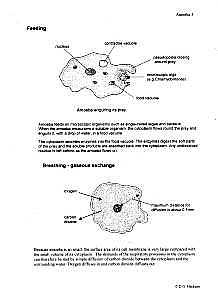
35 Diagram Of Amoeba With Label - Labels Database 2020
Post a Comment for "38 chlamydomonas diagram with labels"